Active
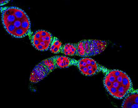
View Entry | Confocal microscopy image of two Drosophila ovarioles5772Ovarioles in female insects are tubes in which egg cells (called oocytes) form at one end and complete their development as they reach the other end of the tube. This image, taken with a confocal microscope, shows ovarioles in a very popular lab animal, the fruit fly Drosophila. The basic structure of ovarioles supports very rapid egg production, with some insects (like termites) producing several thousand eggs per day. Each insect ovary typically contains four to eight ovarioles, but this number varies widely depending on the insect species.
Scientists use insect ovarioles, for example, to study the basic processes that help various insects, including those that cause disease (like some mosquitos and biting flies), reproduce very quickly. | | Public Note | | | | | Internal Note | | Researchers and Olympus granted permission for public use:
Xie, Ting [TGX@stowers.org]
Actions
To:
Spiering, Martin (NIH/NIGMS) [C]
Cc:
Machalek, Alisa Zapp (NIH/NIGMS) [E]
Wednesday, August 03, 2016 7:39 PM
You replied on 8/4/2016 8:32 AM.
Dear Martin,
I am glad that you choose the beautiful image for promotion purpose. Please credit the image in such a way, Daniel Kirilly in the Ting Xie Lab. Please let me know if you need more information on the image. Thanks
Best Regards
Ting
Sent from my iPhone
Spiering, Martin (NIH/NIGMS) [C]
Sent Items
Wednesday, August 03, 2016 3:52 PM
Dear Dr. Xie,
I am a writer and editor with the Office of Communication and Public Liaison at the National Institute of General Medical Sciences. I'm reaching out to you because we would like to include one of your stunning images (showing two fly ovarioles) that was taken by one of your lab associates, Daniel Kirilly, and won honorable mention at the 2004 Olympus BioScapes (at http://www.olympusbioscapes.com/gallery/year/2004) into our image gallery (at https://images.nigms.nih.gov/). We feel the image would greatly enhance our gallery and also bring your work to broader public attention.
Images and videos in the NIGMS image gallery highlight NIGMS-funded work and are made available to the public for educational uses, provided that users credit the creator, i.e., you (or Daniel Kirilly), for this work. Would you let us feature your work in this way? Please note that we have already obtained permission for such use and a high-resolution file of your image from Olympus.
Please let me know if you have any questions or concerns. I look forward to hearing from you.
Thank you,
Martin J Spiering, PhD, ELS
Writer & Editor (contractor)
OCPL, National Institutes of Health/NIGMS
From: Ilene Semiatin [ilene@edge-comm.net]
Sent: Wednesday, August 03, 2016 10:42 AM
To: Spiering, Martin (NIH/NIGMS) [C]
Subject: Re: seeking permission to use Daniel Kirilly image
Hi Martin,
We hereby provide permission to use the 2004 Kirilly image of ovarioles in your gallery provided that you properly credit both Daniel Kirilly and the 2004 Olympus BioScapes Competition in your credit information. Do you need a link to download the image? If so, I can provide it.
Regarding images being in the public domain, some of the images that earn BioScapes awards might be in the public domain by default, as some of them are captured at federal government institutions and some of the research product produced at federally funded institutions is, according to policy, in the public domain. However, we are not the arbiters of what is and what is not in the public domain. Unless a researcher has specifically told us in writing that his/her work is not copyrighted and is in the public domain, we assume that our regular copyright and permissions rules should be adhered to. For your gallery’s purposes, we will be pleased to reply with permission to use any of the images if you agree to credit them properly. Let us know which ones you’d like access to.
Thanks,
Ilene
On Aug 3, 2016, at 10:31 AM, Spiering, Martin (NIH/NIGMS) [C] wrote:
Hello Ilene,
Thank you very much for getting back to me. Strange that my email didn't reach you--I just sent a test email from a different account to see whether this is an issue to do with the originating email system/server.
I have attached the image we like to get permission to include in the gallery. As for your question about why we want to use this image, all media in the NIGMS image gallery highlight NIGMS-funded work and are made available to the public for educational uses, provided that users credit the creator for this work.
As you may recall, my colleague, Alisa Machalek, had asked you about the possibility of these images entering public domain, so I just want to double check this with you. From your note, I understand that all of these images remain copyrighted and won't move into the public domain any time soon. Is that correct?
Please let me know whether we could get permission to include the image in our gallery and if you have any questions or need anything else from us.
Thanks again,
Martin
| | | Keywords | | fruit fly
ovary
Drosophila;
development; developmental biology | | | Source | | 2004 Olympus BioScapes Competition | | | Date | | | | | Credit Line | | Daniel Kirilly in the Ting Xie Lab, Stowers Institute for Medical Research, and the 2004 Olympus BioScapes Competition | | | Investigator | | Ting Xie, Stowers Institute for Medical Research | | | Record Type | | Photograph | | | Topic Area(s) | | ;#Cells;#Tools and Techniques;# | | | Previous Uses | | | | | Status | | Active | |
| | View All Properties | | Edit Properties |
|


