Active
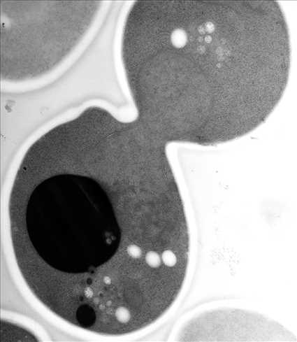
View Entry | EM of yeast cell division5770Cell division is an incredibly coordinated process. It not only ensures that the new cells formed during this event have a full set of chromosomes, but also that they are endowed with all the cellular materials, including proteins, lipids and small functional compartments called organelles, that are required for normal cell activity. This proper apportioning of essential cell ingredients helps each cell get off to a running start.
This image shows an electron microscopy (EM) thin section taken at 10,000x magnification of a dividing yeast cell over-expressing the protein ubiquitin, which is involved in protein degradation and recycling. The picture features mother and daughter endosome accumulations (small organelles with internal vesicles), a darkly stained vacuole and a dividing nucleus in close contact with a cadre of lipid droplets (unstained spherical bodies). Other dynamic events are also visible, such as spindle microtubules in the nucleus and endocytic pits at the plasma membrane.
These extensive details were revealed thanks to a preservation method involving high-pressure freezing, freeze-substitution and Lowicryl HM20 embedding. | | Public Note | | | | | Internal Note | | Researchers gave permission for public use:
From: Matthew C. West [mwest@colorado.edu]
Friday, July 22, 2016 6:25 PM
Martin,
You have my permission to post my EM and tomography models at NIGMS. Best!
--Matt
Matthew West
Odorizzi lab
MCD Biology, University of Colorado, Boulder
303-735-6518
From: Spiering, Martin (NIH/NIGMS) [C]
To: Matthew C. West [mwest@colorado.edu]
Cc:Greg Odorizzi [charles.odorizzi@colorado.edu]
Friday, July 22, 2016 5:14 PM
Hello Matt,
Many thanks for remembering and for sending this great image! I'll add this to our image gallery shortly. Which reminds me, would you give us permission to include these images in the NIGMS image gallery (at https://images.nigms.nih.gov/Pages/Home.aspx)? Images and videos in the NIGMS image gallery highlight NIGMS-funded work and are made available to the public for educational uses, provided that users credit the creator, i.e., you, for this work. Would that be okay?
The post featuring your endosome image should be posted on Biomedical Beat ( https://biobeat.nigms.nih.gov/) shortly--the person who does the posting will be back next week and will hopefully move this forward promptly.
Have a great weekend,
Martin | | | Keywords | | endosome
Saccharomyces cerevisiae
cell division
mitosis | | | Source | | Matthew West and Greg Odorizzi, University of Colorado | | | Date | | | | | Credit Line | | Matthew West and Greg Odorizzi, University of Colorado | | | Investigator | | Greg Odorizzi, University of Colorado | | | Record Type | | Photograph | | | Topic Area(s) | | ;#Cells;#Genes;#Tools and Techniques;# | | | Previous Uses | | | | | Status | | Active | |
| | View All Properties | | Edit Properties |
|


