Active
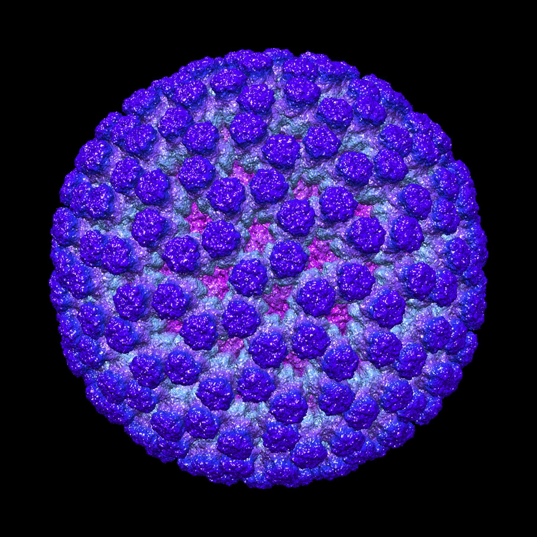
View Entry | Rotavirus structure3584This image shows a computer-generated, three-dimensional map of the rotavirus structure. This virus infects humans and other animals and causes severe diarrhea in infants and young children. By the age of five, almost every child in the world has been infected with this virus at least once. Scientists have found a vaccine against rotavirus, so in the United States there are very few fatalities, but in developing countries and in places where the vaccine is unavailable, this virus is responsible for more than 200,000 deaths each year.
The rotavirus comprises three layers: the outer, middle and inner layers. On infection, the outer layer is removed, leaving behind a "double-layered particle." Researchers have studied the structure of this double-layered particle with a transmission electron microscope. Many images of the virus at a magnification of ~50,000x were acquired, and computational analysis was used to combine the individual particle images into a three-dimensional reconstruction.
The image was rendered by Melody Campbell (PhD student at TSRI). Work that led to the 3D map was published in Campbell et al. Movies of ice-embedded particles enhance resolution in electron cryo-microscopy. Structure. 2012;20(11):1823-8. PMCID: PMC3510009.
This image was part of the Life: Magnified exhibit that ran from June 3, 2014, to January 21, 2015, at Dulles International Airport. | | Public Note | | | | | Internal Note | | From: Bridget Carragher [mailto:bcarr@scripps.edu]
Sent: Thursday, April 24, 2014 7:51 PM
To: Machalek, Alisa Zapp (NIH/NIGMS) [E]
Cc: Gregurick, Susan (NIH/NIGMS) [E]; Smith, Kelley (NIH/NIGMS) [C]; Thea Clarke; Potter Clint; Melody Campbell
Subject: Re: OK to put rotavirus image into public domain?
Yes indeed of course. But if possible can you use the image credit on the web site as follows. This refers to the paper so at least to som extent recognizes the large cast of characters that contributed to the image?.
The image was rendered by Melody Campbell (PhD student at TSRI). Work that led to the 3D map was published in:
Campbell MG, Cheng A, Brilot AF, Moeller A, Lyumkis D, Veesler D, Pan J, Harrison SC, Potter CS, Carragher B, Grigorieff N. Movies of ice-embedded particles enhance resolution in electron cryo-microscopy. Structure. 2012;20(11):1823-8. PMCID: PMC3510009.
[stuff deleted]
On Apr 24, 2014, at 2:33 PM, Machalek, Alisa Zapp (NIH/NIGMS) [E] wrote:
Hi again, Bridget,
Your image is officially in the Dulles exhibit! Would NIGMS also be able to put it into our public domain Image and Video Gallery at http://images.nigms.nih.gov/?
Best,
Alisa | | | Keywords | | Life Magnified, structure, 3d | | | Source | | Bridget Carragher, The Scripps Research Institute, La Jolla, CA | | | Date | | | | | Credit Line | | National Resource for Automated Molecular Microscopy, The Scripps Research Institute | | | Investigator | | Bridget Carragher, The Scripps Research Institute, La Jolla, CA | | | Record Type | | Illustration | | | Topic Area(s) | | ;#Molecular Structures;#Tools and Techniques;# | | | Previous Uses | | Dulles airport exhibit | | | Status | | Active | |
| | View All Properties | | Edit Properties |
|


