Active
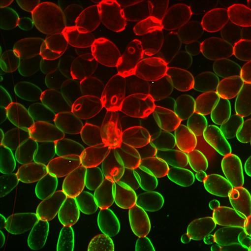
View Entry | Snowflake yeast6969Multicellular yeast called snowflake yeast that researchers created through many generations of directed evolution from unicellular yeast. Stained cell membranes (green) and cell walls (red) reveal the connections between cells. Younger cells take up more cell membrane stain, while older cells take up more cell wall stain, leading to the color differences seen here. This image was captured using spinning disk confocal microscopy.
Related to images 6970 and 6971. | | Public Note | | Many ovals connected to one another. Those at the top of the image are mostly red, while those at the bottom are mostly green, with red spots where they connect to other ovals. | | | Internal Note | | From: Ratcliff, William C william.ratcliff@biology.gatech.edu
Sent: Tuesday, January 3, 2023 4:16 PM
To: Kimberly Rousseau krousseau@iqsolutions.com
Cc: Tony Burnetti tony.burnetti@gmail.com; Burnetti, Anthony J anthony.burnetti@biosci.gatech.edu
Subject: Re: For Review: NIGMS Blog Post
CAUTION: This email originated from an external sender
Hi Kim,
Great! Looking forward to reading the final piece.
I'm more than happy to add these images to the NIGMS gallery! These images have not been used in any papers yet, but I was planning on submitting them as journal cover art. I assume that's still OK if they're in the NIGMS gallery first. If not, I'll tell the journals to shove it. They can't hold copyright over our images.
Credit information: Image by Anthony Burnetti, Ozan Bozdag and Will Ratcliff, Georgia Institute of Techology.
As for the microscopy details, I will let Tony answer this. He is the one who took the pictures.
Tony, can you do me a favor and write a brief title / caption for these images?
Cheers,
Will
ps- Happy New Year!
Associate Professor, Biological Sciences
Director, Interdisciplinary Graduate Program in Quantitative Biosciences (QBioS)
Georgia Institute of Technology
Lab website: http://www.ratclifflab.biology.gatech.edu/
Google Scholar profile
Twitter: @wc_ratcliff
Phone: 612-840-4983
Office: 331 Cherry Emerson
Lab: 330 Cherry Emerson | | | Keywords | | Research organisms, model organisms, saccharomyces cerevisiae, balloons | | | Source | | William Ratcliff, Georgia Institute of Technology. | | | Date | | | | | Credit Line | | Anthony Burnetti, Ozan Bozdağ, and William Ratcliff, Georgia Institute of Technology. | | | Investigator | | “Snowflake yeast” are the result of many generations of settling selection driving yeast into multicellular forms. They consist of branching chains of cells attached to their mothers and daughters. Staining cell membranes (green) and cell walls (red) reveals the connections between the cells, holding them together into a larger whole. The properties of these cell to cell connections change and evolve over continued laboratory selection. Image captured via spinning disk confocal microscopy.
The different colors are coming from the fact that older and younger cells seemingly have a systematically different staining. Younger cells are taking in less cell wall stain, and older cells are systematically taking in less membrane stain. This could be due to the same thing – older cells having more cell wall, which both stains and prevents the membrane stain from getting through as well. | | | Record Type | | Photograph | | | Topic Area(s) | | ;#Cells;#Tools and Techniques;# | | | Previous Uses | | | | | Status | | Active | |
| | View All Properties | | Edit Properties |
|


