Active
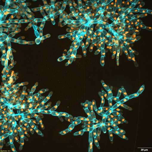
View Entry | Snowflake yeast6971Multicellular yeast called snowflake yeast that researchers created through many generations of directed evolution from unicellular yeast. Here, the researchers visualized nuclei in orange to help them study changes in how the yeast cells divided. Cell walls are shown in blue. This image was captured using spinning disk confocal microscopy.
Related to images 6969 and 6970. | | Public Note | | Several clusters, each made up of many branches of connected blue ovals. Each oval contains an orange circle. | | | Internal Note | | Grant: R35GM138030
From: Ratcliff, William C william.ratcliff@biology.gatech.edu
Sent: Tuesday, January 3, 2023 4:16 PM
To: Kimberly Rousseau krousseau@iqsolutions.com
Cc: Tony Burnetti tony.burnetti@gmail.com; Burnetti, Anthony J anthony.burnetti@biosci.gatech.edu
Subject: Re: For Review: NIGMS Blog Post
CAUTION: This email originated from an external sender
Hi Kim,
Great! Looking forward to reading the final piece.
I'm more than happy to add these images to the NIGMS gallery! These images have not been used in any papers yet, but I was planning on submitting them as journal cover art. I assume that's still OK if they're in the NIGMS gallery first. If not, I'll tell the journals to shove it. They can't hold copyright over our images.
Credit information: Image by Anthony Burnetti, Ozan Bozdag and Will Ratcliff, Georgia Institute of Techology.
As for the microscopy details, I will let Tony answer this. He is the one who took the pictures.
Tony, can you do me a favor and write a brief title / caption for these images?
Cheers,
Will
ps- Happy New Year!
Associate Professor, Biological Sciences
Director, Interdisciplinary Graduate Program in Quantitative Biosciences (QBioS)
Georgia Institute of Technology
Lab website: http://www.ratclifflab.biology.gatech.edu/
Google Scholar profile
Twitter: @wc_ratcliff
Phone: 612-840-4983
Office: 331 Cherry Emerson
Lab: 330 Cherry Emerson | | | Keywords | | Research organisms, model organisms, saccharomyces cerevisiae, nucleus | | | Source | | William Ratcliff, Georgia Institute of Technology. | | | Date | | | | | Credit Line | | Anthony Burnetti, Ozan Bozdağ, and William Ratcliff, Georgia Institute of Technology. | | | Investigator | | Many basic cellular processes are affected by laboratory evolution of multicellular snowflake yeast for large size and increased strength. Pulling apart fragments of multicellular clusters subject to 600 days of evolution for large size, the shape and behavior of individual cells can be observed. Here, fluorescent proteins (orange) localized to nuclei allow changes in the process of cell division to be examined.
The blue is cell wall, a stain called calcofluor, and it is directly staining the polysaccharides. | | | Record Type | | Photograph | | | Topic Area(s) | | ;#Cells;#Tools and Techniques;# | | | Previous Uses | | https://biobeat.nigms.nih.gov/2023/02/career-conversations-qa-with-evolutionary-biologist-william-ratcliff/ | | | Status | | Active | |
| | View All Properties | | Edit Properties |
|


