Active
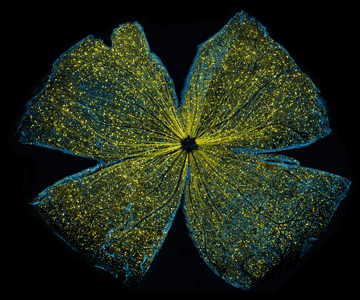
View Entry | Mouse retina5793What looks like the gossamer wings of a butterfly is actually the retina of a mouse, delicately snipped to lay flat and sparkling with fluorescent molecules. The image is from a research project investigating the promise of gene therapy for glaucoma. It was created at an NIGMS-funded advanced microscopy facility that develops technology for imaging across many scales, from whole organisms to cells to individual molecules.
The ability to obtain high-resolution imaging of tissue as large as whole mouse retinas was made possible by a technique called large-scale mosaic confocal microscopy, which was pioneered by the NIGMS-funded National Center for Microscopy and Imaging Research. The technique is similar to Google Earth in that it computationally stitches together many small, high-resolution images.
| | Public Note | | | | | Internal Note | | From: Sammak, Paul (NIH/NIGMS) [E]
Sent: Thursday, October 13, 2016 12:53 PM
To: Ellisman Mark (mark@ncmir.ucsd.edu) ; 'deerinck@ncmir.ucsd.edu' ; 'keunyoung@ncmir.ucsd.edu' ; 'wju@ucsd.edu'
Cc: Machalek, Alisa Zapp (NIH/NIGMS) [E] ; Gregurick, Susan (NIH/NIGMS) [E] ; Greenberg, Judith (NIH/NIGMS) [E] ; Sheeley, Douglas (NIH/NIGMS) [E]
Subject: NIH image recognition: Lighting Up the Promise of Gene Therapy for Glaucoma.
Dear Christine, Tom, Wonkyu and Mark,
Your image of GFP-labeled ganglia in a mouse retina preparation was selected for first prize in the Combined Federal Campaign NIH IC Directors Art Challenge for “The Beauty of Science” (http://cfc.nih.gov/). It was selected by combined voting of IC Directors for the best three images. The wining art will be displayed in Building 1, the CFC website, and in upcoming NIH publications.
I also would like to acknowledge the elegant work in the paper that describes the encouraging story about the promise of therapy for Glaucoma from basic science studies. Please share with your co-authors!
PAPER:
DRP1 inhibition rescues retinal ganglion cells and their
axons by preserving mitochondrial integrity in a mouse
model of glaucoma
K-Y Kim1, GA Perkins1, MS Shim2, E Bushong1, N Alcasid1, S Ju1, MH Ellisman1, RN Weinreb2 and W-K Ju*,2
Citation: Cell Death and Disease (2015) 6, e1839; doi:10.1038/cddis.2015.180
& 2015 Macmillan Publishers Limited All rights reserved 2041-4889/15
www.nature.com/cddis
More details:
Glaucoma is a progressive eye disease and the leading cause of irreversible blindness. It is characterized by the death of neurons in the retina called retinal ganglion cells. A number of studies over the past decade suggest that targeting these cells with gene therapy designed to prevent their death might slow the progression of glaucoma.
This study is investigating whether a non-disease-causing virus (adeno-associated virus serotype 2) can effectively deliver genes to retinal ganglion cells. The researchers introduced into the virus a gene for green fluorescent protein (GFP) so they could visualize how well the virus transduced the cells.
Two months after viral delivery of the fluorescent vector to the eyes of 7-month-old mice, the researchers examined the entire retinas of the subjects under a microscope. The ability to obtain high-resolution imaging of tissue as large as whole mouse retinas was made possible by a technique called large-scale mosaic confocal microscopy, which was pioneered by the NIGMS-funded National Center for Microscopy and Imaging Research. The technique is similar to Google Earth in that it computationally stitches together many small, high-resolution images.
The researchers observed GFP expression (yellow) in all parts of the retinal ganglion cells (blue), including the soma, axons and dendritic tree. These results suggest that a viral delivery system could deliver therapeutic genes to retinal ganglion cells for treating glaucoma and related diseases.
EQUIPMENT: Olympus FluoView™ FV1000 Confocal Microscope. Fluorophores: green fluorescent protein and Alexa Fluor 568. Non-glaucomatous DBA/2J-Gpnmb+ mice.
Reflecting on the work, the lead researcher [Keunyoung (“Christine”) Kim] says: “It is amazing to see intricate and artistically organized microscopic structures. … I encountered an entirely new world invisible to the naked eye—a galaxy of infinite secrets and endless potential for discovery.”
| | | Keywords | | retina
large-scale mosaic confocal microscopy
CFC
| | | Source | | Tom Deerinck and Keunyoung (“Christine”) Kim, NCMIR | | | Date | | | | | Credit Line | | Keunyoung Kim, Wonkyu Ju and Mark Ellisman, National Center for Microscopy and Imaging Research, University of California, San Diego. | | | Investigator | | Keunyoung (“Christine”) Kim, Wonkyu Ju and Mark Ellisman, all of the National Center for Microscopy and Imaging Research (NCMIR) at the University of California, San Diego.
Grant info: R24GM137200 | | | Record Type | | Photograph | | | Topic Area(s) | | ;#Cells;#Tools and Techniques;# | | | Previous Uses | | BioBeat; won first place in the 2016 CFC NIH Director's art contest
| | | Status | | Active | |
| | View All Properties | | Edit Properties |
|


