Active
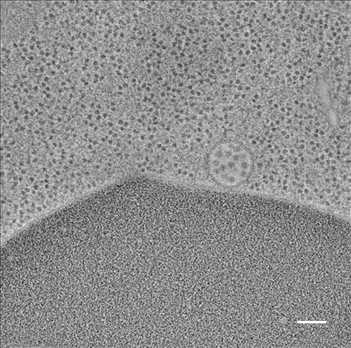
View Entry | Multivesicular bodies containing intralumenal vesicles assemble at the vacuole 25768Collecting and transporting cellular waste and sorting it into recylable and nonrecylable pieces is a complex business in the cell. One key player in that process is the endosome, which helps collect, sort and transport worn-out or leftover proteins with the help of a protein assembly called the endosomal sorting complexes for transport (or ESCRT for short). These complexes help package proteins marked for breakdown into intralumenal vesicles, which, in turn, are enclosed in multivesicular bodies for transport to the places where the proteins are recycled or dumped. In this image, a multivesicular body (the round structure slightly to the right of center) contain tiny intralumenal vesicles (with a diameter of only 25 nanometers; the round specks inside the larger round structure) adjacent to the cell's vacuole (below the multivesicular body, shown in darker and more uniform gray).
Scientists working with baker's yeast (Saccharomyces cerevisiae) study the budding inward of the limiting membrane (green lines on top of the yellow lines) into the intralumenal vesicles. This tomogram was shot with a Tecnai F-20 high-energy electron microscope, at 29,000x magnification, with a 0.7-nm pixel, ~4-nm resolution.
To learn more about endosomes, see the Biomedical Beat blog post The Cell’s Mailroom. Related to a color-enhanced version 5767 and image 5769. | | Public Note | | | | | Internal Note | | Researchers gave permission for public use:
From: Matthew C. West [mwest@colorado.edu]
Friday, July 22, 2016 6:25 PM
Martin,
You have my permission to post my EM and tomography models at NIGMS. Best!
--Matt
Matthew West
Odorizzi lab
MCD Biology, University of Colorado, Boulder
303-735-6518
From: Spiering, Martin (NIH/NIGMS) [C]
To: Matthew C. West [mwest@colorado.edu]
Cc:Greg Odorizzi [charles.odorizzi@colorado.edu]
Friday, July 22, 2016 5:14 PM
Hello Matt,
Many thanks for remembering and for sending this great image! I'll add this to our image gallery shortly. Which reminds me, would you give us permission to include these images in the NIGMS image gallery (at https://images.nigms.nih.gov/Pages/Home.aspx)? Images and videos in the NIGMS image gallery highlight NIGMS-funded work and are made available to the public for educational uses, provided that users credit the creator, i.e., you, for this work. Would that be okay?
The post featuring your endosome image should be posted on Biomedical Beat ( https://biobeat.nigms.nih.gov/) shortly--the person who does the posting will be back next week and will hopefully move this forward promptly.
Have a great weekend,
Martin
| | | Keywords | | | | | Source | | Matthew West and Greg Odorizzi, University of Colorado | | | Date | | | | | Credit Line | | Matthew West and Greg Odorizzi, University of Colorado | | | Investigator | | Greg Odorizzi, University of Colorado | | | Record Type | | Photograph | | | Topic Area(s) | | ;#Cells;#Tools and Techniques;# | | | Previous Uses | | vacuole
endocytosis
endosome
recycling | | | Status | | Active | |
| | View All Properties | | Edit Properties |
|


