Active
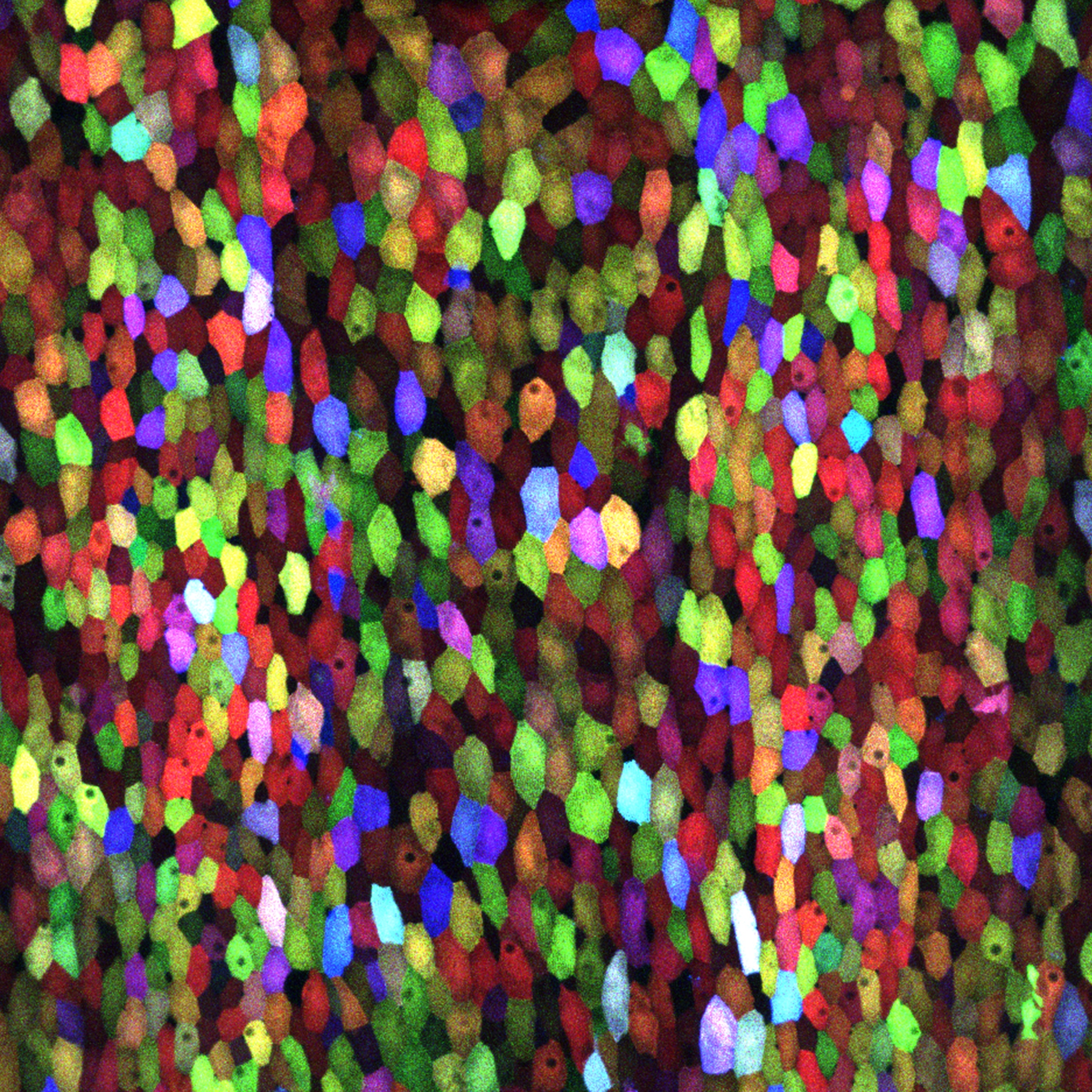
View Entry | A multicolored fish scale 13782Each of the colored specs in this image is a cell on the surface of a fish scale. To better understand how wounds heal, scientists have inserted genes that make cells brightly glow in different colors into the skin cells of zebrafish, a fish often used in laboratory research. The colors enable the researchers to track each individual cell, for example, as it moves to the location of a cut or scrape over the course of several days. These technicolor fish endowed with glowing skin cells dubbed "skinbow" provide important insight into how tissues recover and regenerate after an injury.
For more information on skinbow fish, see the Biomedical Beat blog post Visualizing Skin Regeneration in Real Time and a press release from Duke University highlighting this research. Related to image 3783. | | Public Note | | | | | Internal Note | | Researchers sent the image an gave permission for public use (image has not been published, see below):
From: Ken Poss, Ph.D. [kenneth.poss@duke.edu]
To: Spiering, Martin (NIH/NIGMS) [C]
Cc: Chen-Hui Chen, Ph.D. [chen-hui.chen@duke.edu]
Monday, May 09, 2016 1:35 PM
Hi Martin,
Chen-Hui Chen has told me that the images now attached are similar to, but areNOT the images published by Developmental Cell.
Hope these will be fine.
Best,
Ken
From: Ken Poss, Ph.D. [kenneth.poss@duke.edu]
Sent: Thursday, March 24, 2016 2:57 PM
To: Spiering, Martin (NIH/NIGMS) [C]
Subject: Re: Skinbow image for NIGMS Image Gallery?
Dear Martin,
Thanks for noticing our work. Yes this is fine with me. I am not sure what you would need from the journal, however. I?ve attached a higher-res image. The credit is Chen-Hui Chen, Developmental Cell 2016.
Best,
Ken Poss
Kenneth Poss
James B. Duke Professor
Duke University Medical Center
Durham, NC 27710
On Mar 24, 2016, at 2:06 PM, Spiering, Martin (NIH/NIGMS) [C] wrote:
Dear Dr. Poss,
I am a writer and editor with the Office of Communication and Public Liaison at the National Institute of General Medical Sciences. I am reaching out to you because we noticed your stunning image of multicolored fish skin cells in this recent news release (http://today.duke.edu/2016/03/zebrafish ) describing your intriguing color-based technique to track individual cells. We would be very grateful if we could include your artwork in our Image Gallery on the NIGMS website (at https://images.nigms.nih.gov/).
Images and videos in the NIGMS Image Gallery highlight NIGMS-funded work and are made available to the public for educational uses, provided that users credit the creator, i.e., you, for this work. Would you let us feature your work in our image gallery in this way? | | | Keywords | | | | | Source | | Chen-Hui Chen and Kenneth Poss, Duke University | | | Date | | | | | Credit Line | | Chen-Hui Chen and Kenneth Poss, Duke University | | | Investigator | | Kenneth Poss, Duke University | | | Record Type | | Photograph | | | Topic Area(s) | | ;#Cells;#Genes;# | | | Previous Uses | | | | | Status | | Active | |
| | View All Properties | | Edit Properties |
|


