Active
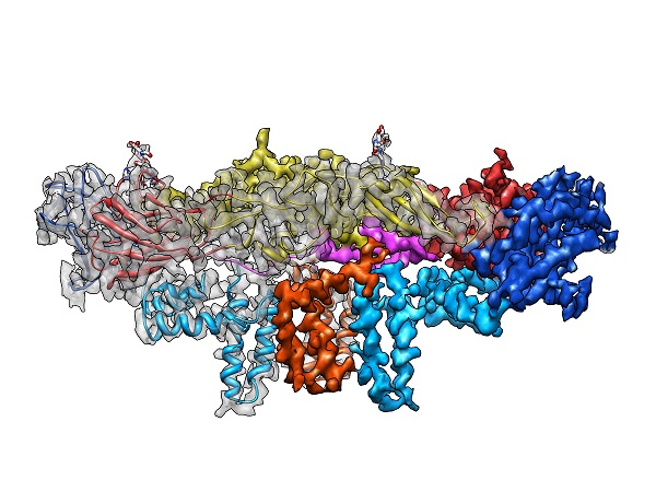
View Entry | Dengue virus membrane protein structure3758| Dengue virus is a mosquito-borne illness that infects millions of people in the tropics and subtropics each year. Like many viruses, dengue is enclosed by a protective membrane. The proteins that span this membrane play an important role in the life cycle of the virus.
Scientists used cryo-EM to determine the structure of a dengue virus at a 3.5-angstrom resolution to reveal how the membrane proteins undergo major structural changes as the virus matures and infects a host. The image shows a side view of the structure of a protein composed of two smaller proteins, called E and M. Each E and M contributes two molecules to the overall protein structure (called a heterotetramer), which is important for assembling and holding together the viral membrane, i.e., the shell that surrounds the genetic material of the dengue virus. The dengue protein's structure has revealed some portions in the protein that might be good targets for developing medications that could be used to combat dengue virus infections.
For more on cryo-EM see the blog post Cryo-Electron Microscopy Reveals Molecules in Ever Greater Detail. You can watch a rotating view of the dengue virus surface structure in video 3748. | | Public Note | | | | | Internal Note | | Researcher gave permission for public use:
Dear Martin,
Please find a high resolution still image in the attachment. I also attach a side view of the protein subunits that make up the virus.
Thank you and have a wonderful weekend!
Best regards,
Hong
Z. Hong Zhou, Ph.D., 310.983.1033/206.0033 Email: Hong.Zhou@UCLA.edu
Electron Imaging Center for Nanomachines (EICN), CNSI http://www.EICN.ucla.edu
and Dept of Microbiology, Immun. & Mol. Genetics http://www.mimg.ucla.edu/
UCLA, Los Angeles, CA 90095-7364 | Office: CNSI 6350A, cell: 310-694-7527
UCLA Mail code: 736422, BSRB 237
Courier delivery: 570 Westwood Plaza, UCLA Building 114, CNSI 6350, Los Angeles, CA 90095
Spiering, Martin (NIH/NIGMS) [C]
To:
Hong.Zhou@UCLA.edu
Cc:
Machalek, Alisa Zapp (NIH/NIGMS) [E]
Sent Items
Wednesday, March 09, 2016 4:03 PM
You replied on 3/11/2016 7:20 AM.
Dear Dr. Zhou,
I am a writer and editor with the Office of Communication and Public Liaison at the National Institute of General Medical Sciences. As you may recall, a colleague of mine, Carolyn Beans, had been in touch with you about the video of the dengue virus structure you had determined by cryo-EM and which is now also posted on our website (at https://images.nigms.nih.gov/index.cfm?event=viewDetail&imageID=3748). Thank you again for letting us feature your great work!
I just have a follow-up question: would you have a high-resolution still image of the dengue virus cryo-EM? we usually like to include also a still image of the videos we feature, and a high-res image would be very ideal. Please let me know if you have any questions.
Thank you,
Martin J Spiering, PhD, ELS
Writer & Editor (contractor)
OCPL, National Institutes of Health/NIGMS
martin.spiering@nih.gov | | | Keywords | | cryo-electron microscopy | | | Source | | Hong Zhou, UCLA | | | Date | | 2016-03-16 00:00:00 | | | Credit Line | | Hong Zhou, UCLA | | | Investigator | | Hong Zhou, UCLA | | | Record Type | | Illustration | | | Topic Area(s) | | ;#Molecular Structures;#Tools and Techniques;# | | | Previous Uses | | | | | Status | | Active | |
| | View All Properties | | Edit Properties |
|
