Active
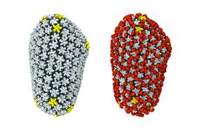
View Entry | Cryo-EM reveals how the HIV capsid attaches to a human protein to evade immune detection3755| The illustration shows the capsid of human immunodeficiency virus (HIV) whose molecular features were resolved with cryo-electron microscopy (cryo-EM). On the left, the HIV capsid is "naked," a state in which it would be easily detected by and removed from cells. However, as shown on the right, when the viral capsid binds to and is covered with a host protein, called cyclophilin A (shown in red), it evades detection and enters and invades the human cell to use it to establish an infection.
To learn more about how cyclophilin A helps HIV infect cells and how scientists used cryo-EM to find out the mechanism by which the HIV capsid attaches to cyclophilin A, see this news release by the University of Illinois. A study reporting these findings was published in the journal Nature Communications. | | Public Note | | | | | Internal Note | | Researchers gave permission for public use:
From: jrperillaj@gmail.com [jrperillaj@gmail.com] on behalf of Juan R. Perilla [jperilla@illinois.edu]
Sent: Monday, March 07, 2016 11:16 AM
To: Spiering, Martin (NIH/NIGMS) [C]
Cc: Schulten, Klaus J
Subject: Re: Inclusion of HIV-cyclophilin A image in NIGMS image gallery
Dear Martin,
I would be happy if you include my figure in NIH's image repository.
You can download a high version of the figure using the following
link:
https://www.dropbox.com/s/y12v3za0t8ezk2s/CaCypA-sidebyside.tif?dl=0
Best regards,
~ Juan
On Mon, Mar 7, 2016 at 9:58 AM, Spiering, Martin (NIH/NIGMS) [C]
wrote:
Dear Dr. Schulten,
I am a writer and editor with the Office of Communication and Public Liaison
at the National Institute of General Medical Sciences. I am reaching out to
you because we just noticed one of your very striking illustrations (see
attached) depicting HIV covered in cyclophilin A featured in this recent
release (https://news.illinois.edu/blog/view/6367/335013) about your
exciting structural biology work on HIV. We would be very grateful if we
could include your artwork in our Image Gallery on the NIGMS website (at
https://images.nigms.nih.gov/).
As you may know, images and videos in the NIGMS Image Gallery highlight
NIGMS-funded work and are made available to the public for educational uses,
provided that users credit the creator, i.e., you, for this work. Would you
let us feature your work in our image gallery in this way? If so, could you
send us a high-resolution version of your illustration (if available)?
Please let me know if you have any questions.
I look forward to hearing from you.
Thank you,
Martin J Spiering, PhD, ELS
Writer & Editor (contractor)
OCPL, National Institutes of Health/NIGMS
martin.spiering@nih.gov | | | Keywords | | | | | Source | | Juan R. Perilla, University of Illinois at Urbana-Champaign | | | Date | | | | | Credit Line | | Juan R. Perilla, Klaus Schulten and the Theoretical and Computational Biophysics Group | | | Investigator | | Klaus Schulten, University of Illinois at Urbana-Champaign | | | Record Type | | Illustration | | | Topic Area(s) | | ;#Molecular Structures;#Tools and Techniques;# | | | Previous Uses | | | | | Status | | Active | |
| | View All Properties | | Edit Properties |
|


