Active
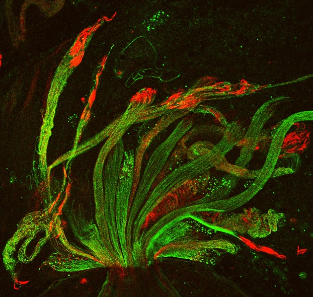
View Entry | Fruit fly spermatids3590| Developing spermatids (precursors of mature sperm cells) begin as small, round cells and mature into long-tailed, tadpole-shaped ones. In the sperm cell's head is the cell nucleus; in its tail is the power to outswim thousands of competitors to fertilize an egg. As seen in this microscopy image, fruit fly spermatids start out as groups of interconnected cells. A small lipid molecule called PIP2 helps spermatids tell their heads from their tails. Here, PIP2 (red) marks the nuclei and a cell skeleton-building protein called tubulin (green) marks the tails. When PIP2 levels are too low, some spermatids get mixed up and grow with their heads at the wrong end. Because sperm development is similar across species, studies in fruit flies could help researchers understand male infertility in humans. | | Public Note | | | | | Internal Note | | -----Original Message-----
From: Lacramioara Fabian [mailto:lala.fabian@utoronto.ca]
Sent: Wednesday, December 11, 2013 5:47 PM
To: Machalek, Alisa Zapp (NIH/NIGMS) [E]
Cc: fuller@cmgm.stanford.edu; Ben-Ari, Elia (NIH/NIGMS) [C]; Carlson, Emily (NIH/NIGMS) [E]; julie.brill@sickkids.ca
Subject: Re: permission for NIH to use your 2010 cover image?
Dear Alisa,
Thank you for selecting my cover image for all of these very exciting publications and events that will help spread the beauty of science.
I am very happy to give you permission to use it in all the mentioned cases.
I am cross copying Dr. Julie Brill on this email. She is my supervisor and also the corresponding author on the MBoC paper.
Thank you again and good luck with your projects.
Best wishes,
Lacramioara
-----------------------
Lacramioara Lala Fabian, PhD
Research Associate
Hospital for Sick Children
Department of Cell Biology
Quoting "Machalek, Alisa Zapp (NIH/NIGMS) [E]" :
> Dear Drs. Fabian and Fuller,
> My office at the U.S. National Institutes of Health would like to
> feature the cover image (attached) from your 2010 MBoC
> paper. With your
> permission, we hope to include the image in an online article for
> our Inside Life
> Science
> series. Articles in the Inside Life Science series also appear on
> the commercial LiveScience.com website.
>
> Do we have permission to use your image for Inside Life Science and
> post it on LiveScience.com?
>
> You may be wondering why we're asking about an image that was
> published a few years ago. The image was one of a dozen or so
> recently selected by staff at the American Society of Cell Biology
> for its beauty. Together, ASCB and NIH's National Institute of
> General Medical Sciences are collecting stunning sc | | | Keywords | | fertility, structure, Grant 2R01GM062276 | | | Source | | Lacramioara Fabian, The Hospital for Sick Children, Toronto, Canada | | | Date | | 2014-05-02 00:00:00 | | | Credit Line | | Lacramioara Fabian and Julie Brill, The Hospital for Sick Children, Toronto, Canada | | | Investigator | | Lacramioara Fabian and Julie Brill
Cover art: https://www.molbiolcell.org/toc/mboc/21/9
Confocal micrograph of Drosophila
melanogaster spermatids. The
spermatids develop in highly
polarized syncytial cysts that
grow to a length of nearly 2 mm.
Here phosphatidylinositol
4,5-bisphosphate (PIP2; red) marks
the nuclei, and polyglycylated
tubulin (green) marks the sperm
tails. In addition to its nuclear
localization, PIP2 also localizes to
the growing ends of spermatid cysts
(not shown) and is required for
spermatid cyst polarity. See the
article by Fabian et al. on p. 1546
of this issue of MBoC. (Image:
Lacramioara Fabian, The Hospital
for Sick Children, Toronto, Ontario,
Canada; anti–polyglycylated tubulin
antibody courtesy of Martin
Gorovsky, University of Rochester) | | | Record Type | | Photograph | | | Topic Area(s) | | ;#Cells;#Genes;#Molecular Structures;#Tools and Techniques;# | | | Previous Uses | | | | | Status | | Active | |
| | View All Properties | | Edit Properties |
|


