Active
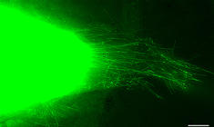
View Entry | Cytonemes in developing fruit fly cells3574| Scientists have long known that multicellular organisms use biological molecules produced by one cell and sensed by another to transmit messages that, for instance, guide proper development of organs and tissues. But it's been a puzzle as to how molecules dumped out into the fluid-filled spaces between cells can precisely home in on their targets.
Using living tissue from fruit flies, a team led by Thomas Kornberg of the University of California, San Francisco, has shown that typical cells in animals can talk to each other via long, thin cell extensions called cytonemes (Latin for "cell threads") that may span the length of 50 or 100 cells. The point of contact between a cytoneme and its target cell acts as a communications bridge between the two cells.
More information about the research behind this image can be found in a Biomedical Beat Blog posting from February 2014. | | Public Note | | | | | Internal Note | | From: Ben-Ari, Elia (NIH/NIGMS) [C]
Sent: Monday, February 17, 2014 4:53 PM
To: Reynolds, Sharon (NIH/NIGMS) [C]
Cc: Machalek, Alisa Zapp (NIH/NIGMS) [E]
Subject: FW: Cytoneme image
Hi Sharon and Alisa,
Attached is an image showing threadlike cytonemes in the developing tracheal system (air sacs, to be specific) of a fruit fly. We have permission from Dr. Thomas Kornbeg of UCSF (see yellow highlighting below) to use it in our image library (and for my Feb. BioBeat item). The cytonemes carry signals between cells in the developing fruit fly, similar to the way neurons convey signals in the nervous system.
The credit for this micrograph goes to Sougata Roy, also of UCSF. The plasma membranes and cytonemes are labeled with green fluorescent protein.
Let me know if you need further info/help in developing a lay language description. I’ve pasted the text of my BioBeat item below, which may help.
Elia
Scientists have long known that multicellular organisms use biological molecules produced by one cell and sensed by another to transmit messages that, for instance, guide proper development of organs and tissues. But it’s been a puzzle as to how molecules dumped out into the fluid-filled spaces between cells can precisely home in on their targets.
Using living tissue from fruit flies, a team led by Thomas Kornberg of the University of California, San Francisco, has shown that typical cells in animals can talk to each other via long, thin cell extensions called cytonemes (Latin for “cell thread[s]”) that may span the length of 50 or 100 cells. The point of contact between a cytoneme and its target cell acts as a communications bridge between the two cells.
Until now, only nerve cells (neurons) were known to communicate this way. “This is an exciting finding,” says NIGMS’ Tanya Hoodbhoy. “Neurons are not the only ‘reach out and touch someone’ cells.”
From: Kornberg, Tom [mailto:Tom.Kornberg@ucsf.edu]
Sent: Friday, February 14, 2014 2:00 PM
To: Ben-Ari, Elia (NIH/NIGMS) [C]
Subject: Re: Cytoneme image
It helps to actually attach it!
--
Thomas Kornberg
Cardiovascular Research Institute
555 Mission Bay South, Room 252z
University of California
San Francisco, CA 94158
Phone: 415-476-8821
From: "Ben-Ari, Elia (NIH/NIGMS) [C]"
Date: Fri, 14 Feb 2014 18:56:43 +0000
To: "Kornberg, Tom"
Subject: RE: Cytoneme image
Many thanks, Tom. The attachment didn’t come through, though.
Elia
From: Kornberg, Tom [mailto:Tom.Kornberg@ucsf.edu]
Sent: Friday, February 14, 2014 1:37 PM
To: Ben-Ari, Elia (NIH/NIGMS) [C]
Subject: Re: Cytoneme image
Elia-
Please see attached figure of an Air Sac Primordium that expresses CD8:GFP that marks its plasma membranes and cytonemes. Micrograph taken by Sougata Roy. You may use it for your purposes.
Tom
--
Thomas Kornberg
Cardiovascular Research Institute
555 Mission Bay South, Room 252z
University of California
San Francisco, CA 94158
Phone: 415-476-8821
From: "Ben-Ari, Elia (NIH/NIGMS) [C]"
Date: Fri, 14 Feb 2014 16:21:50 +0000
To: "Kornberg, Tom"
Subject: Cytoneme image
Hello Tom,
My understanding, after checking with a colleague, is that if AAAS holds the copyright on the figure it will also apply to the photo without the arrows and lettering.
Do you have a photo/illustration of a cytoneme (or cytonemes) that you retain the rights to? And if so, may we use it for the purposes I outlined below?
Thanks again for your help.
Elia
From: Kornberg, Tom [mailto:Tom.Kornberg@ucsf.edu]
Sent: Wednesday, February 12, 2014 5:13 PM
To: Ben-Ari, Elia (NIH/NIGMS) [C]
Subject: Re: Please review: Short piece on your recent work for NIGMS news blog
You certainly have my permission, but should probably assume that AAAS will hold a copyright on the figure as they will publish it with the arrows and lettering. Does their copyright still apply if you use it without the arrows and lettering?
--
Thomas Kornberg
Cardiovascular Research Institute
555 Mission Bay South, Room 252z
University of California
San Francisco, CA 94158
Phone: 415-476-8821
From: "Ben-Ari, Elia (NIH/NIGMS) [C]"
Date: Wed, 12 Feb 2014 22:03:40 +0000
To: "Kornberg, Tom"
Subject: RE: Please review: Short piece on your recent work for NIGMS news blog
Thanks very much, Tom.
Does AAAS hold a copyright on the figure? If so, I’d need to get their permission to use it. Also, who should be credited for the image?
If you retain the copyright, do you grant NIGMS permission to use it as part of our Biomedical Beat blog? And would you also be willing to grant us permission to use the image in our image gallery, provided that users give proper credit? (Images in the gallery could be used for educational, news media, or research purposes.) If not, no problem.
Apologies for all the questions.
Elia
From: Kornberg, Tom [mailto:Tom.Kornberg@ucsf.edu]
Sent: Wednesday, February 12, 2014 4:50 PM
To: Ben-Ari, Elia (NIH/NIGMS) [C]
Subject: Re: Please review: Short piece on your recent work for NIGMS news blog
Elia-
Your summary is fine. I've attached two versions of a figure (with and without arrows&lettering) and a legend. The figure will appear in the print version of Science, that will be a one page summary (also attached). The full article will be published online only.
Best,
Tom
--
Thomas Kornberg
Cardiovascular Research Institute
555 Mission Bay South, Room 252z
University of California
San Francisco, CA 94158
Phone: 415-476-8821
From: "Ben-Ari, Elia (NIH/NIGMS) [C]"
Date: Wed, 12 Feb 2014 19:58:06 +0000
To: "tkornberg@ucsf.edu"
Subject: Please review: Short piece on your recent work for NIGMS news blog
Dear Dr. Kornberg:
I’m writing to let you know that we’ve featured a link to the news release on your recent Science paper on cytonemes on the NIGMS Web site. In addition, we plan to include a brief summary of your advance on our research news blog, Biomedical Beat. Could you please review the attached draft and send me any comments by Friday, Feb. 14?
We’d like to publish a relevant image with the summary. Is there a nice, clear image of cytonemes that we might use with the story that you could send along? Perhaps one of the panels from a figure in theScience paper (minus the arrows)? Please also send a brief description of the image let me know who should be credited, as we include photo credits (name, affiliation) in these blog items.
If you provide an image, please let me know in writing (email is fine) that you grant us permission to feature the image on our blog. In addition, we would love to include it in our collection of research-related images. Images in this gallery are made available for educational, news media, or research purposes, provided that users credit the source of the image, i.e., you or whomever you indicate. If you grant us permission to feature the image in our gallery, please send a high-resolution version.
In the future, we would very much like to work with you and the press officers at your institution to help publicize the results of NIGMS support, so please let us know when you have another manuscript accepted for publication that describes a significant finding we funded. We will, of course, honor all embargoes on journal articles. Contact us at 301-496-7301 or info@nigms.nih.gov.
Thanks for your help!
Best regards,
Elia Ben-Ari
---------
Elia Ben-Ari, PhD
Science writer (contractor)
Office of Communications and Public Liaison
National Institute of General Medical Sciences
National Institutes of Health
elia.ben-ari@nih.gov
OCPL main number: 301-496-7301
| | | Keywords | | strcture | | | Source | | Sougata Roy, University of California, San Francisco | | | Date | | | | | Credit Line | | Sougata Roy, University of California, San Francisco | | | Investigator | | Thomas Kornberg , University of California, San Francisco, | | | Record Type | | Photograph | | | Topic Area(s) | | ;#Cells;#Molecular Structures;#Tools and Techniques;# | | | Previous Uses | | | | | Status | | Active | |
| | View All Properties | | Edit Properties |
|
