Active
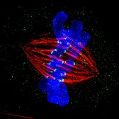
View Entry | Dividing cell in metaphase3445This image of a mammalian epithelial cell, captured in metaphase, was the winning image in the high- and super-resolution microscopy category of the 2012 GE Healthcare Life Sciences Cell Imaging Competition. The image shows microtubules (red), kinetochores (green) and DNA (blue). The DNA is fixed in the process of being moved along the microtubules that form the structure of the spindle.
The image was taken using the DeltaVision OMX imaging system, affectionately known as the "OMG" microscope, and was displayed on the NBC screen in New York's Times Square during the weekend of April 20-21, 2013. It was also part of the Life: Magnified exhibit that ran from June 3, 2014, to January 21, 2015, at Dulles International Airport. | | Public Note | | | | | Internal Note | | Hi Alisa,
It was also great to hear from you as well. It has been so many years.
I've attached the TIF image, which is higher resolution than the jpg image.
I have other images, but they are all over the place right now and not in an easy condensed format. They are also of very recent data that is not yet published so we haven't decided which ones we will use for publication yet and which are the extras.
Claire
Hi Claire,
I hope that no one you know was affected by the Boston bombings yesterday. How awful!
It was so nice to chat with you the other day and I'm delighted to learn that you?re on the ASCB outreach committee. Please tell the group that I'd be happy to partner with them to help educate the public about the importance of basic research.
I finally got permission from GE to put your OMG image into our public domain image gallery. Just checking--is the one posted as "print-quality" on http://newsinfo.iu.edu/pub/libs/images/usr/15146_h.jpg the highest resolution version you have?
I'd also love to feature other pretty pictures from you, so feel free to send me some (free of any copyright restrictions) if you get a chance. We have a blog, a small Pinterest site and other outlets that would be great ways to highlight your work.
Keep it up!
Alisa
From: Rebecca Caygill [mailto:Rebecca.Caygill@collegehill.com]
Hi Alisa,
Thanks you for your email and sorry for the delay in responding.
GE Healthcare Life Sciences have confirmed that you have permission to share Jane's image but ask that the NIH credit is as detailed below:
Metaphase epithelial cell in metaphase stained for microtubules (red), kinetochores (green) and DNA (blue). Jane Stout, Indiana University, GE Healthcare 2012 Cell Imaging Competition.
With best wishes,
Rebecca | | | Keywords | | Cell division, cell cycle, imaging, cell imaging, mitosis | | | Source | | Jane Stout in the laboratory of Claire Walczak, Indiana University, GE Healthcare 2012 Cell Imaging Competition | | | Date | | 2013-04-16 00:00:00 | | | Credit Line | | Jane Stout and Claire Walczak, Indiana University | | | Investigator | | | | | Record Type | | Photograph | | | Topic Area(s) | | ;#Cells;#Genes;# | | | Previous Uses | | Dulles exhibit 2014 | | | Status | | Active | |
| | View All Properties | | Edit Properties |
|
